Patello-Femoral Joint
The patella-femoral joint (PFJ) is a complex articulation between the patella and the distal femoral condyle. 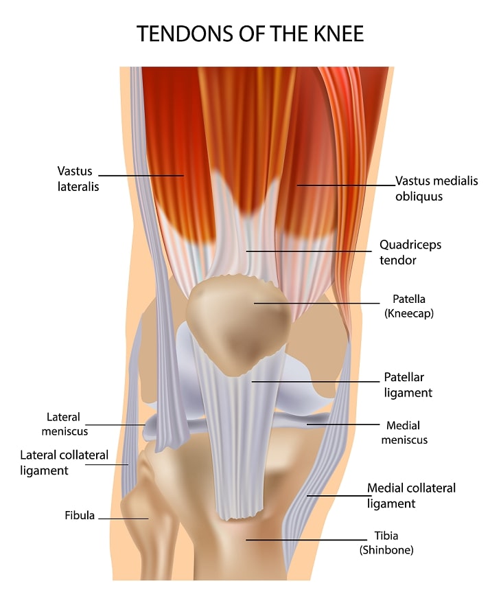
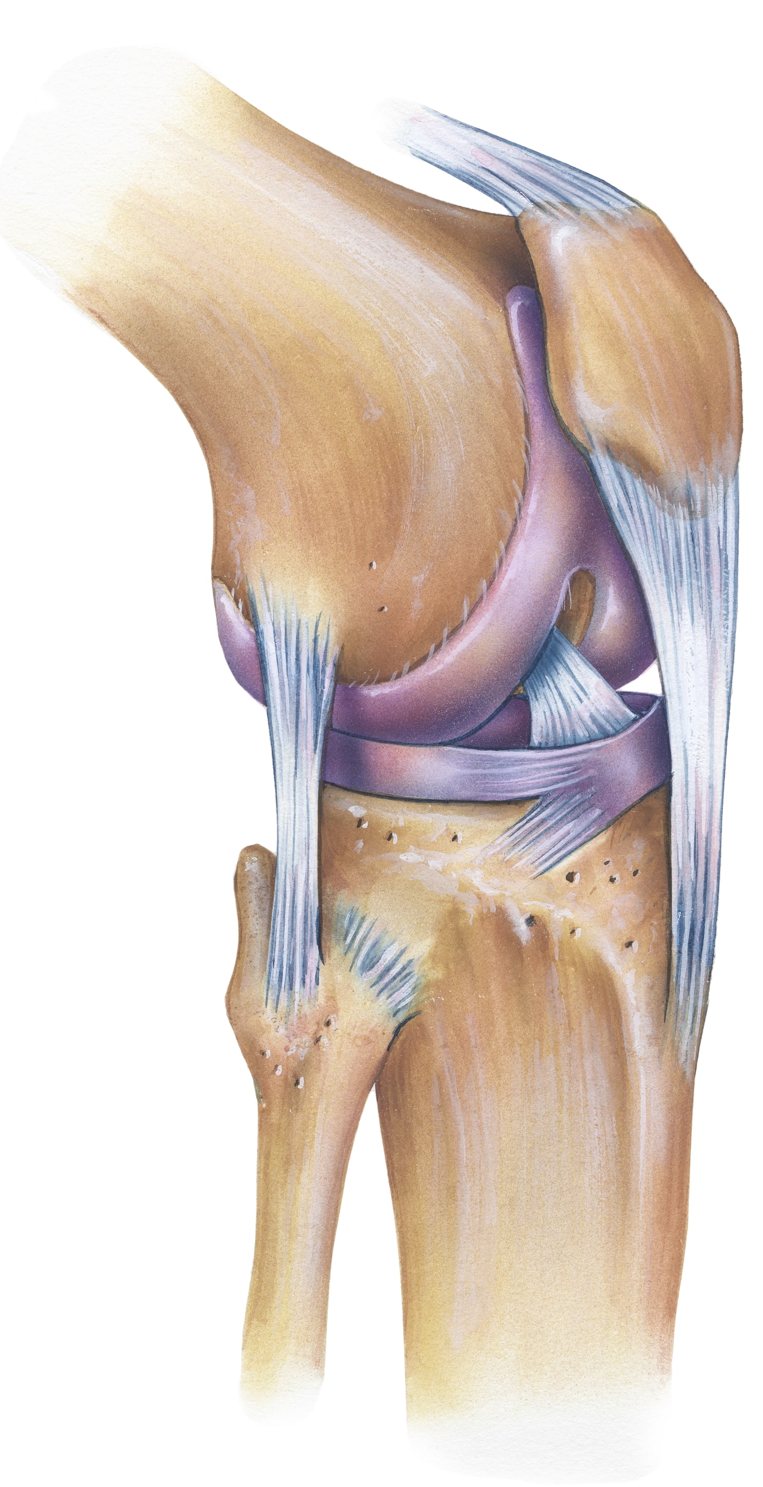 The shape of the femur and patella provides bony constraint once the patella engages into the trochlea groove when knee flexion exceeds 30 degrees. The medial patella-femoral ligament (MPFL) provides static stability and is the primary restraint from 0-20 degrees of flexion.
Patellar Instability
Patellar instability can be categorized into acute and traumatic, chronic laxity and habitual laxity. 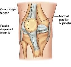 Traumatic laxity may occur via a direct blow or by a non-contact twisting injury with the knee extended and externally rotated. Acute dislocations are associated with large effusions, obvious deformity and pain. The absence of swelling indicates chronic and habitual conditions. One complication that is frequently seen is damage to the articular cartilage on the medial patella facet. If there is no loose body in the knee non-operative treatment is the first line of treatment. Loose bodies require keyhole surgery and removal. Treatment of chronic patellar instability is complex and many factors must be taken not account. An important factor is overall alignment of the lower extremity and may require corrective bone surgery. If there is no underlying malalignment repair or reconstruction of the MPFL is a successful procedure.
Lateral Patellar Compression Syndrome
If the patella does not track properly in the trochlea groove there is pain with stairclimbing and when sitting for prolonged periods of time. It is caused by tight lateral structures and leads to excessive tilt of the patella.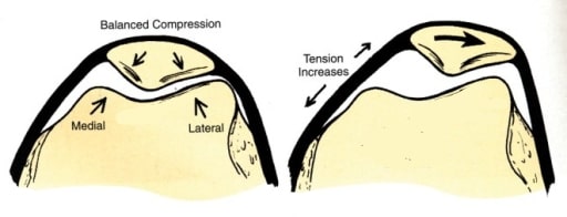 Pain on compression and tenderness over the lateral facet of the patella are clinical signs. Everting the lateral edge of the patella is difficult. Non-operative is the mainstay of treatment. If there is no improvement with conservative measures surgery may be a good option. If there is no malalignment and patellar instability release of the lateral structures can be performed with keyhole surgery.
Idiopathic Chondromalacia Patellae
This condition is characterized by changes in the articular cartilage of the patella. 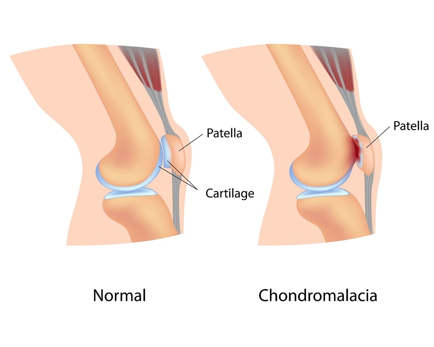 It occurs most commonly in young adolescents and adults and is more prevalent in females. Common symptoms are diffuse pain in the region of the patella. It is insidious in onset and is aggravated by daily activities but especially with climbing or descending stairs, prolonged sitting with the knee bend and squatting or kneeling. If there are no deformities or malignment the mainstay of treatment is rest, rehabilitation and anti-inflammatories. Steroid injections are not helpful. Arthroscopic debridement is indicated with higher grade changes.
Quadriceps Tendon Rupture
A complete tear of the quadriceps tendon usually occurs in people older than 40 years. In most cases the tendon tears at the insertion into the patella. 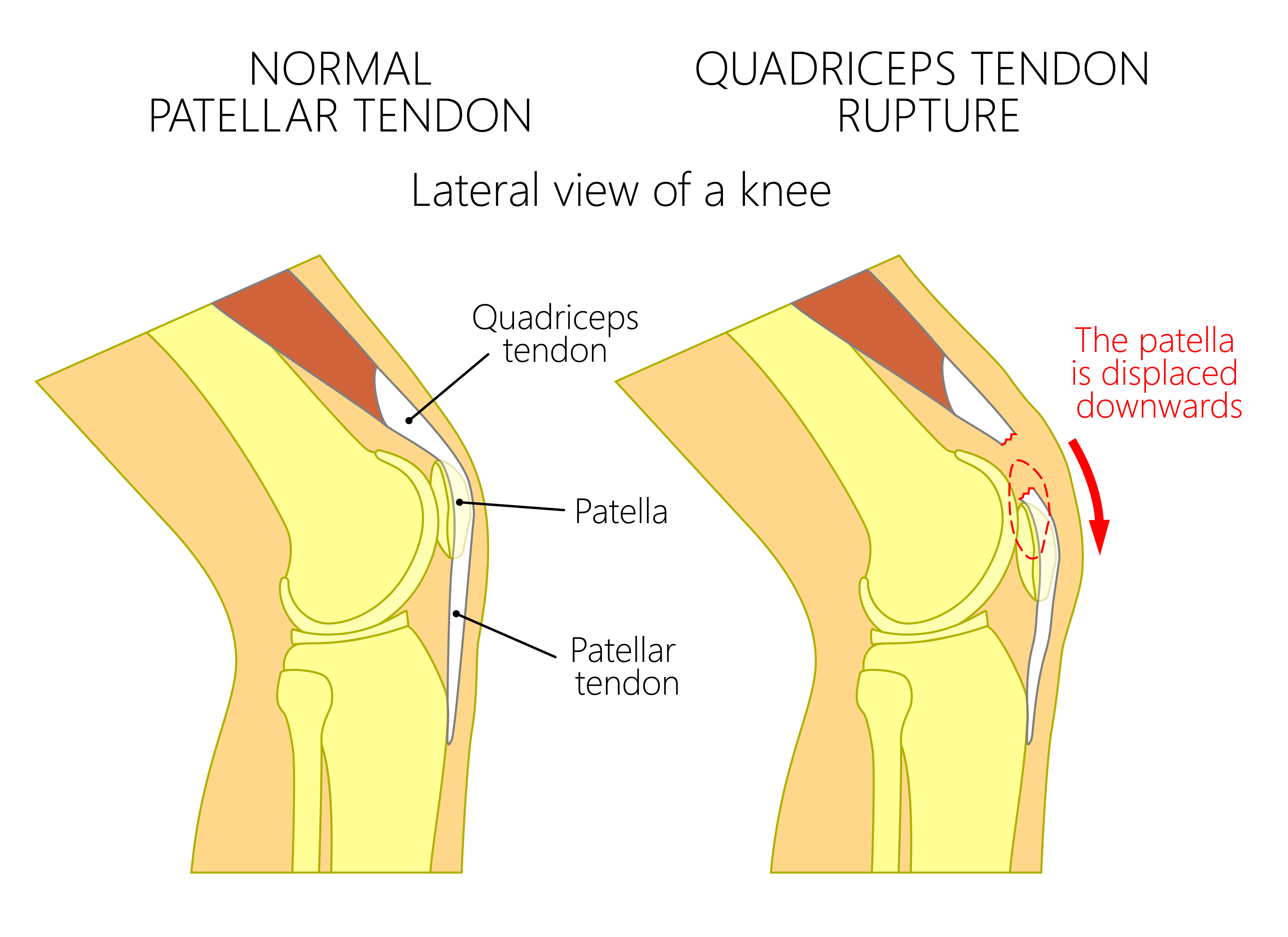 Eccentric loading with the foot slightly bent and direct trauma are the described mechanisms of injury. Complete tears result in inability to extend the knee and extreme difficulties in walking. Surgery is almost always indicated. Strength deficits, stiffness and functional impairment following surgery are not uncommon.
Patella Tendon Rupture
A complete tear of the patella tendon occurs in the younger population. Most ruptures occur with the knee flexed. Risk factors include any condition which weakens the collagen structure: systemic lupus erythematous, rheumatoid arthritis, chronic renal disease and diabetes mellitus.
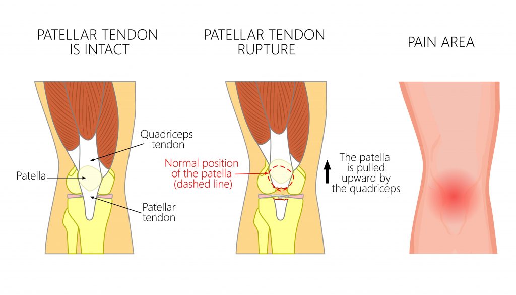 Other factors include degenerative changes of the patella tendon, patella tendinitis, previous injury and cortisone injections. Nonoperative treatment can be entertained with partial tears. Surgical treatment is normally indicated and severely disrupted or degenerate tendons often require augmentation with hamstring grafts. Atrophy of the quadriceps muscle, stiffness and loss of knee flexion and decreased knee extension strength are reported complications
